Allow 30 to 45 minutes. Sometimes an examination or an urgent patient takes a little more time; thank you for your understanding if it occasionally takes longer.
Radiologie YDB BV (Radiologie Buggenhout), Hauwerstraat 40, 9255 Buggenhout, is responsible for processing your personal data. Questions? Email info@radiologiebuggenhout.be or call 052 577 227.
Patients
We process your identity and contact details, your health insurance details and your medical data (request, images and report).
We do this to perform your examination, send the result to your doctors and bill the examination. This is necessary for your care and required by law.
Your data only goes to care providers treating you (referring doctor, GP, via CoZo and eHealth), to your health insurer, and to the companies that securely store images and reports for us. They work on our behalf under strict agreements.
We keep your medical record as long as the law requires: at least 30 years.
Applicants
We use your name, contact details, motivation and CV only for your application and keep them for at most one year.
Website
This website uses no cookies and does not track your visit.
Online appointments and image viewing run through external services (Cheduler and PacsOnWeb), each with its own privacy statement.
Your rights
You can view your data, have it corrected or, where the law allows, erased. You can also object to processing. Email info@radiologiebuggenhout.be. If you are not satisfied, you can lodge a complaint with the Data Protection Authority, Drukpersstraat 35, 1000 Brussels.
Last updated: October 2026
What is it? An X-ray image. A small dose of X-rays passes through the body. Bones block the rays and appear white on the image, while soft tissues let them through.
What is it for? Fractures, osteoarthritis, rheumatic conditions, spinal alignment, pneumonia.
What happens? You stand, sit or lie still while the image is taken. No preparation is needed. If you are or might be pregnant, please tell us beforehand.
What is it? Imaging with sound waves, without radiation. A probe with a little gel on the skin shows live images of what lies beneath. Doppler also shows the blood flow.
What is it for? Tendons, muscles and joints, swellings and lumps, abdominal organs such as the liver, gallbladder and kidneys, the thyroid, blood vessels.
What happens? You lie comfortably while the radiologist performs the examination personally. It is painless. An abdominal or pelvic ultrasound sometimes needs preparation (see Preparation).
What is it? An ultrasound of the blood vessels, without radiation. Doppler and duplex show not only the vessel itself, but also measure how fast and in which direction the blood flows.
What is it for?
Neck vessels: narrowing or calcification of the carotid arteries
Vessels of the arms and legs: arteries and veins, for example in narrowing, thrombosis or varicose veins
Aorta: widening or narrowing of the body’s main artery
Renal vessels: narrowing of the kidney arteries
What happens? You lie comfortably while the radiologist examines the vessels with a probe and a little gel. It is painless.
What is it? A low-dose X-ray examination of the breasts. With 3D tomosynthesis the breasts are imaged in thin layers, so small abnormalities are easier to see. Depending on the density of the breast tissue or your medical history, an additional ultrasound is strongly recommended. The two examinations often complement each other.
What is it for? Breast cancer screening (we are an accredited screening centre), examining a palpable lump or other symptoms, and follow-up.
What happens? The breast is briefly compressed between two plates in 2 directions. The pressure can be uncomfortable, but it is really necessary for a good examination. It only lasts a few seconds each time. Do not use deodorant, powder or body lotion that day. Bring previous breast images if they were taken elsewhere. If needed, an image-guided biopsy can be done.
What is it? A CT scanner designed specifically to make very thin slices. Bone is therefore more clearly visible than on a conventional CT.
What is it for? Face and sinuses, teeth and jaw, the inner ear, and joints such as the wrist, hand, knee and ankle, possibly with contrast in the joint (arthrography).
Dental: pre-implant examinations, including pseudo-panoramic images (pseudo-OPG).
What happens? You lie still on the table while the scanner rotates around the area being examined. It takes less than a minute.
For an arthrography the radiologist first injects contrast into the joint under local anaesthesia. Please come with someone who can drive you home afterwards.
What is it? A measurement of bone density (DEXA) with a very low dose of X-rays. The result shows how strong your bones are.
What is it for? Detecting and monitoring osteoporosis, and estimating your risk of fractures. Often requested after the menopause or with long-term use of cortisone.
What happens? You lie on a table while an arm moves over your hip and lower back. It is painless. Do not take calcium supplements in the 24 hours before. Undissolved calcium tablets in the gut can make the result falsely high.
What is it? An X-ray examination in which you drink a contrast liquid so that the oesophagus and stomach show up on the images. We use Gastrografin, a clear water-soluble contrast agent.
What is it for?
Swallow study: checking how you swallow, for example if you choke or feel that food gets stuck
Oesophagus study: narrowing, pouches, acid reflux or a hiatal hernia
Oesophagus and stomach study: oesophagus, stomach and the start of the small intestine, for example with stomach complaints or after stomach surgery
What happens? Do not eat for 4 hours beforehand. You drink a few sips of contrast while the radiologist takes live images, standing and sometimes lying down. You swallow each time the radiologist asks.
After the examination Gastrografin tastes a little bitter and can temporarily loosen your stools. Drink enough water that day. You can eat and go about your normal activities straight away.
Are you pregnant or allergic to iodinated contrast? Please let us know beforehand.
Performed on a high-quality radiological scanner and assessed by a certified radiologist.
What is it? A measurement of your full body composition with the same scanner we use for bone densitometry, a GE Lunar Prodigy. We measure fat mass, muscle mass and body fat percentage, for the whole body and per region (arms, legs and trunk).
What is it for? For athletes, to monitor training and nutrition, but also during a diet or rehabilitation. By repeating the scan after some time, we can track your progress accurately.
What happens? You lie still on a table while an arm moves over your body. It is painless and the radiation dose is very low.
Please note: this examination is not reimbursed by RIZIV/INAMI or your health insurance fund.
Price: €60
What is it? A treatment in which the radiologist injects a product or PRP exactly in the right place. The needle is followed on the image the whole time, usually with ultrasound, so the product or PRP ends up precisely where it is needed.
What is it for? Pain from osteoarthritis, tendinitis, bursitis, trigger finger and carpal tunnel syndrome.
Corticosteroids: reduce inflammation and pain
Hyaluronic acid: lubricates the joint in osteoarthritis
Lumbar facet injection: treatment of the small joints in the lower back
Hydrodilatation: under radiographic guidance, for adhesive capsulitis (frozen shoulder)
What happens? The injection is done after disinfection and under local anaesthesia. It may be somewhat painful for the first 2 days after treatment. Please come with someone who can drive you home afterwards.
What is it? PRP stands for platelet-rich plasma. Your blood is centrifuged in a tube specially designed for this purpose, concentrating your own platelets and growth factors. These are then injected under ultrasound guidance into the affected tendon, after the radiologist has carefully prepared the tendon.
What is it for? Healing of inflamed or torn tendons, such as tennis elbow, golfer’s elbow, Achilles tendinitis or plantar fasciitis (heel spur).
What happens? We take a little blood and prepare the PRP. After disinfection, the procedure is done under local anaesthesia, without affecting the treatment itself. The radiologist works under continuous ultrasound guidance. Nevertheless, it is often a painful procedure that can be clearly felt for up to 2 weeks. Please come with someone who can drive you home afterwards.
Do not take anti-inflammatories (such as Voltaren, Brufen or Apranax) from 2 weeks before to 2 weeks after the treatment.
What is it? An ultrasound-guided treatment of hydroxyapatite calcifications, mainly in the shoulder, using a specific single- or dual-plane needle technique. The calcium deposit is punctured, broken up and rinsed out.
We have more than 10 years of experience with this technique.
What is it for? A painful calcium deposit in a tendon (calcific tendinitis), usually in the shoulder.
What happens? After disinfection and a local anaesthetic, the radiologist performs the treatment under continuous ultrasound guidance. It may be somewhat painful for the first 2 days after treatment. Please come with someone who can drive you home afterwards.
What is it? With a fine needle we take a small piece of tissue or some cells. The needle is followed on the image the whole time, so we hit exactly the right spot. The sample goes to the lab for analysis.
What is it for? To find out exactly what an abnormality is, such as a lump in the breast or thyroid, an enlarged lymph node, or a swelling in the soft tissues (under the skin or in a muscle).
Aspiration and drainage: an abscess or fluid collection is punctured under imaging guidance and aspirated or drained.
What happens? The biopsy is done after disinfection and under local anaesthesia. You get a small plaster. The area may be a little sore for a few days. Please come with someone who can drive you home afterwards. Do you take blood thinners? Ask your GP whether you need to stop them temporarily; it depends on the medicine. The result goes to your doctor.
Sports radiologyInjectionsHead and neckNeuroradiology
Coordinator Radiologie Buggenhout
Radiologist since 2015 (VUB)
Fellowships at the University of Florida (2015) and KU Leuven (2016)
President of VERB-AREB 2020–2025
Former international instructor in musculoskeletal ultrasound and head and neck radiology courses, including in South Africa
Former lecturer in head and neck radiology at Odisee Brussels and VUB (medicine)
Ultrasound- and X-ray-guided injections
Facet joint injections
PRP injections for tendon disorders and osteoarthritis
Has been performing barbotage for more than 10 years with his own single- and dual-plane needle techniques
Our practice is accredited and not bound by the official fee agreement. In addition to the official fee, we may charge a supplement that is not reimbursed by your health insurance fund.
The amount differs per examination. Feel free to ask at reception or by phone before the examination starts.
Consultants: Dr Pakeezah De Beul, Dr Julie Desimpel.
Your doctor’s referral form. Any form is valid: ours, the official RIZIV/INAMI form or one from another hospital. The referral must come from a doctor or dentist; a referral from a physiotherapist or osteopath is not sufficient. Without a referral we cannot perform the examination; this is a legal requirement.
Your ID card or a sticker from your health insurance fund.
Upper abdominal ultrasound (liver, gallbladder, pancreas): fast for 4 hours beforehand. Do not eat; a little water is fine, but no coffee, no fizzy drinks such as cola and no smoking.
Swallow study or upper GI study: fast for 4 hours beforehand, as above.
Ultrasound of the kidneys or pelvis (uterus, ovaries, bladder, prostate): drink about 1 litre of water in the hour before the examination and do not empty your bladder.
Mammography: no deodorant, powder or body lotion. Bring previous breast images if they were taken elsewhere.
Bone densitometry: no calcium supplements in the 24 hours before. Undissolved calcium tablets in the gut can make the result falsely high.
Fine-needle aspiration or biopsy: do you take blood thinners? Ask your GP whether you need to stop them temporarily.
PRP: no anti-inflammatories from 2 weeks before to 2 weeks after the treatment.
Injection, barbotage, aspiration, biopsy or arthrography: come with someone who can drive you home afterwards.
Other examinations need no preparation.
You only pay the patient contribution and any supplement. Most of the cost is paid directly by your health insurance fund.
Bancontact, Bancontact app, Payconiq or cash
No credit cards
After 18:00: €10 surcharge
Missed appointment without notice: €25
More than 30 minutes late without notice: €10
Can’t make it? Let us know as soon as possible on 052 577 227, so we can help another patient sooner.
The report is written right after the examination. You receive a card with a reference number starting with NK9.
With that number and your date of birth you can view the report and images on PacsOnWeb. Your referring doctor and GP receive the result electronically, usually within the hour. Any doctor treating you can also view the report and images via CoZo after reading your ID card.
You can also give the code to your physiotherapist, so they can view your images and report.
Doctors and dentists
Login: your e-mail address, with your Dobcomed password
Forgot your password? 03 780 17 40 or support@dobcomed.com
Forward to colleagues with the Forward button, top right
Images stay online for at least 2 years, after that on request
E-mail a referral: info@radiologiebuggenhout.be
Physiotherapists
Physiotherapists cannot request an examination themselves, but we work closely with them.
The patient receives a card with a code (NK9…). With that code and the patient’s date of birth, the physiotherapist can view the images and report online, to assess the treatment or refer the patient back to the doctor.
For questions about an examination or a patient’s follow-up, you can always contact us.

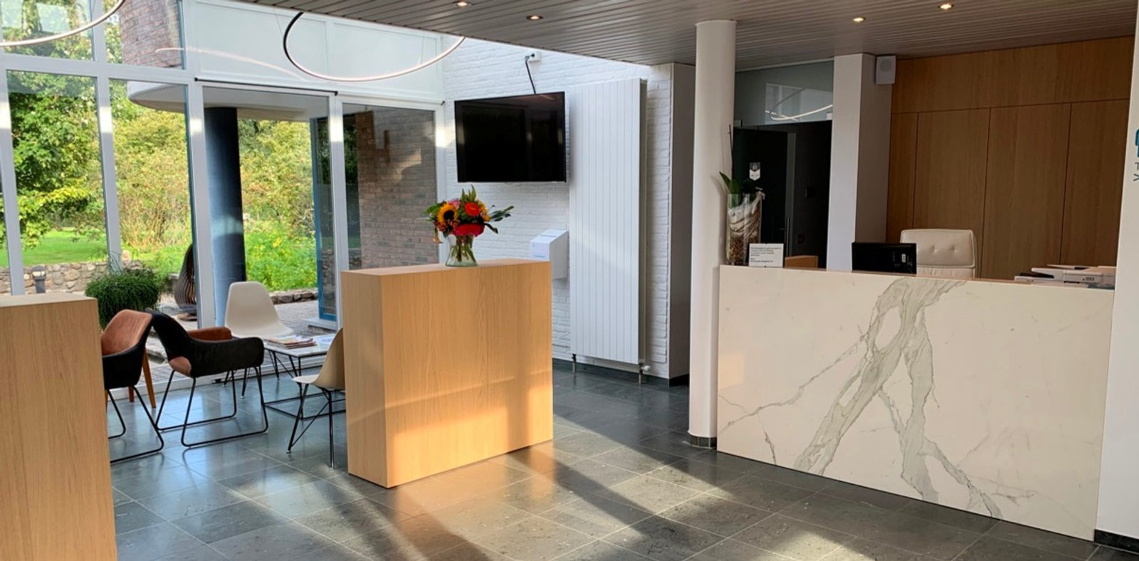 Our reception